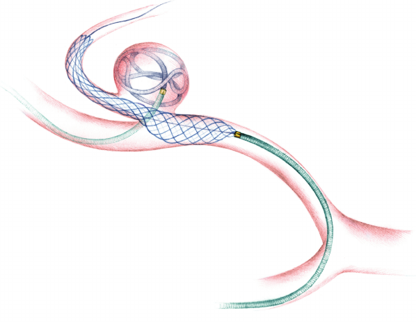
An-illustration-showing-the-LEO-Baby-stent-assisted-coiling-treatment-of-a-PICA-aneurysm

First Brain Aneurysm Coiling in Goa

Interventional Procedure

Brain Aneurysm Coiling

Procedure Date

February 2018

In the Media

https://timesofindia.indiatimes.com/city/goa/docs-perform-brain-aneurysm-coiling/articleshow/46292111.cms

Introduction

brain aneurysm, also known as a subarachnoid hemorrhage (SAH), is a weak spot in the wall of a blood vessel inside the brain

That area of the blood vessel gets worn out from the constant flow of blood and bulges out, almost like a bubble. It can grow to the size of a small berry. It has the risk of rupturing and leading to bleeding in the brain cavity.

Diagnosis & Treatement

A 42-year-old Russian was complaining of severe headache, vomiting and loss of consciousness.

On further evaluation of the brain arteries with CT angiography, it was found that the right posterior inferior cerebellar artery (PICA), a small artery in the posterior portion of the brain, had a ruptured aneurysm arising from its wall.
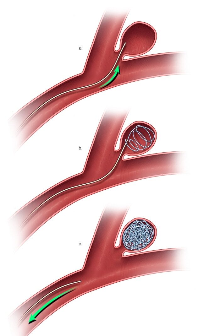
As an emergency, the patient was taken up for aneurysm coiling”. The aneurysm coiling, also called endovascular coiling, was performed by Dr. Charudutt Sambhaji and Dr. Keerthiraj, both vascular and interventional radiologists at Manipal hospital Goa in a procedure that lasted for around two-and-a-half hours.


When an aneurysm ruptures it results in a catastrophic brain hemorrhage. Ten to 15% of these patients die before reaching the hospital, with more than 50% of fatalities occurring in the first 30 days after rupture. Of those who survive, approximately half suffer some permanent neurological deficit. PICA aneurysms, the type identified in the patient is among the rare and technically difficult aneurysms to treat by coiling 

Conclusion

In a first of its kind, rare procedure was performed to save the life of a patient suffering from a brain aneurysm and internal hemorrhage.  The patient recovered successfully without any complications.


Varicose Veins

Varicose Veins Treatment: Traditional Surgery Vs Laser

Interventional Procedure

Endovenous Laser Surgery

Introduction

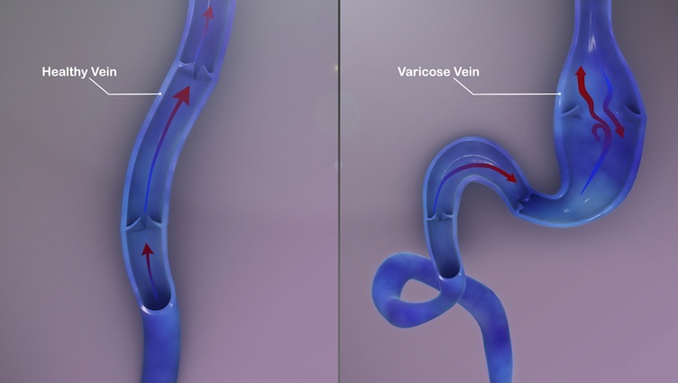
Varicose veins are twisted, enlarged veins close to the skin’s surface (superficial). Varicose veins are caused by increased blood pressure in the veins. 

In the human body, blood moves towards the heart by one-way valves in the veins. When the valves become weakened or damaged, blood can collect in the veins. This causes the veins to become enlarged. Sitting or standing for long periods can cause blood to pool in the leg veins, increasing the pressure within the veins causing stretching of the veins &  weakening of the walls.

Symptoms & Diagnosis

Varicose veins might not always cause pain. Signs of varicose veins include:

  • Veins that are dark purple or blue
  • Veins that appear twisted and bulging, often appearing like cords on the legs

When painful signs and symptoms of varicose veins occur, they might include:

  • An achy or heavy feeling in the legs
  • Burning, throbbing, muscle cramping & swelling in the lower legs
  • Worsened pain after sitting or standing for a long time
  • Itching around one or more of the veins
  • Changes in skin color around a varicose vein


Treatment

The various medical treatments for Varicose veins include 

  • Compression stockings: These elastic stockings squeeze the veins and prevent blood from pooling. Compression stockings can be effective if they are worn every day.

  • Traditional SurgerY: Ligation and stripping are the most commonsurgical interventions for the treatment of varicose veins. Ligation is the process involving surgical tying off of a large vein in the leg while stripping is the process of removal of the same vein via incisions or tissue cuts in the groin region, thigh and leg.
  • Foam Sclerotherapy: Sclerotherapy is the most common treatment for both spider and varicose veins. A salt (saline) or chemical solution is injected into the varicose veins. They no longer carry blood. And, other veins take over.

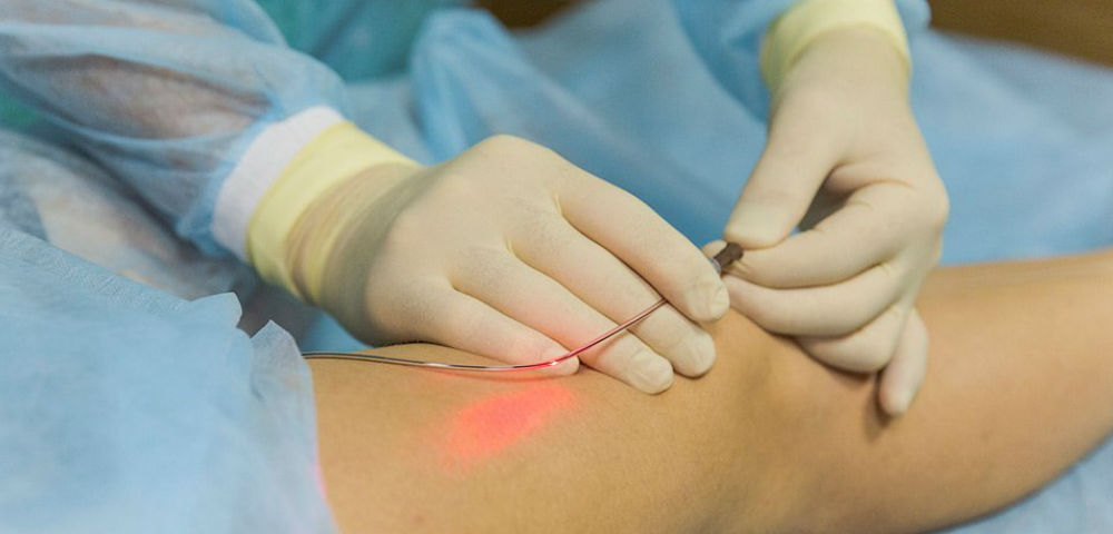
  • Endovascular Laser Abalation Therapy (EVLT):  Lasers or radiofrequency energy may be used to treat varicose veins. A tiny fiber is inserted into a varicose vein through a catheter. The laser or radiofrequency energy is used to deliver heat that destroys the wall of the varicose vein.

  • Ambulatory phlebectomy: Special tools inserted through small cuts (incisions) are used to remove varicose veins. It may be done alone or with vein stripping.

Complcations of Surgical Treatment

Surgical interventions which are undertaken under general anesthesia usually the 1- 2 hours depending upon the no incisions and complications. Moreover, the recovery period is also long from 2-4 weeks depending upon factors like patient’s health & the number of operated veins.

Other complications which could arise are 

  • Allergic reaction to the numbing solution or local anesthetic that’s used.
  • Infection at or near the incision site.
  • Nerve injury can cause long-term numbness near the treatment site.
  • Heavy bleeding.
  • Pronounced scars.
  • Blood clots.
  • Injury to the vein or surrounding tissues.

Advantages of Endovasular Laser Abalation Therapy

Unlike surgical treatments, Endovascular laser therapy(EVLT)  is performed under local anesthesia.. It is comparatively a safer procedure associated with low risks, lesser pain or discomfort, and a higher success rate that is around 98%.

It requires comparatively lesser time for procedure and patients are able to go back home walking on the same day.

Moreover, patients require lesser post-operative care even at home. The patient can resume his normal activities almost immediately which is much earlier than surgical interventions.

Conclusion

Interventional radiology guided minimal or pinhole access procedures simplify the treatment of Varicose veins by using an innovative approach to treat them, causing minimal discomfort, side effects & complications & a much less recovery period.

Please consult your doctor if you have the symptoms & signs  associated with Varicose Veins 

Uterine fibroids

Uterine Fibroid Embolization – Is it the right procedure for you?

Interventional Procedure

Uterine Fibroid Embolization

Introduction

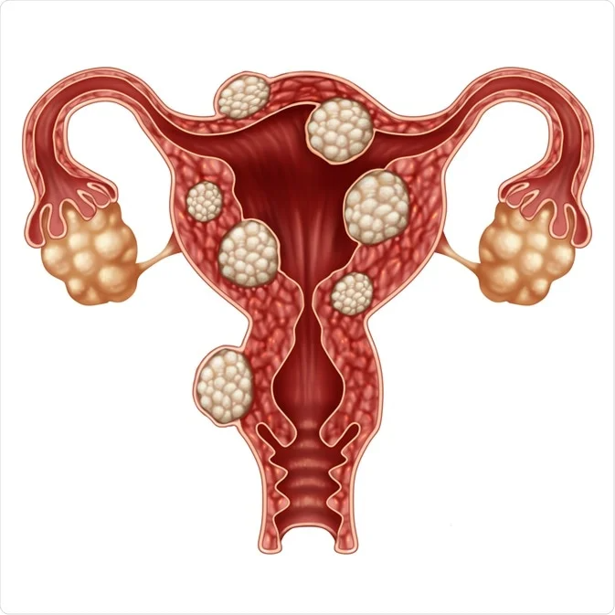
Fibroid tumors are noncancerous (benign) growths that develop in the muscular wall of the uterus. While fibroids do not always cause symptoms, their size and location can lead to problems for some women, including pain and heavy bleeding. Fibroids can range in size from very tiny to the size of small fruit. In some cases, they can cause the uterus to grow to the size of a five-month pregnancy or more.

Symptoms & Diagnosis

Many women who have fibroids don’t have any symptoms. In those that do, symptoms can be influenced by the location, size, and number of fibroids.

In women who have symptoms, the most common signs and symptoms of uterine fibroids include:

  • Heavy menstrual bleeding
  • Menstrual periods lasting more than a week
  • Pelvic pressure or pain
  • Frequent urination
  • Difficulty emptying the bladder
  • Constipation
  • Backache or leg pains


Treatment

Traditional surgical procedures

Options for traditional surgical procedures include:

  • Abdominal myomectomy. If you have multiple fibroids, very large fibroids or very deep fibroids, your doctor may use an open abdominal surgical procedure to remove the fibroids.

    Many women who are told that hysterectomy is their only option can have an abdominal myomectomy instead. However, scarring after surgery can affect future fertility.

  • Hysterectomy. This surgery removes the uterus. It remains the only proven permanent solution for uterine fibroids.

    Hysterectomy ends your ability to bear children. If you also elect to have your ovaries removed, the surgery brings on menopause and the question of whether you’ll take hormone replacement therapy. Most women with uterine fibroids may be able to choose to keep their ovaries.

Alternatives to Surgical Treatment & Hystrectomy

Women suffering from painful uterine fibroids — the most common cause of hysterectomies — now have an alternative, fibroid embolization, which can save them from having the surgical procedure. Interventional radiologists at The Johns Hopkins Hospital perform the fibroid embolization procedure, which involves cutting off blood flow to the fibroid, causing it to shrink.

What is Uterine Fibroid Embolization (UFE) ?


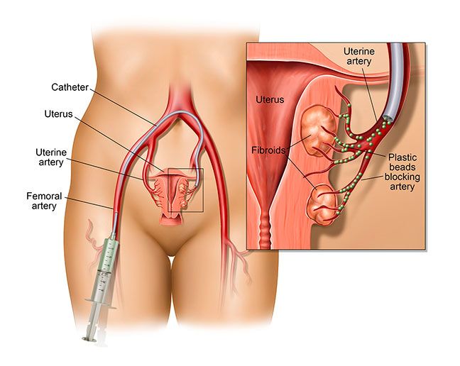
Fibroid embolization, also called uterine artery embolization, is done without surgery. It requires only a tiny nick in the skin about the size of a pencil tip. It is performed while the patient is conscious but sedated — drowsy and feeling no pain. Fibroid embolization is performed by an interventional radiologist, a physician who is specially trained to perform this and other minimally invasive procedures.

The interventional radiologist makes a small nick in the skin in the groin, through which a tiny tube called a catheter is inserted into an artery. The catheter is guided through the artery to the uterus while the interventional radiologist watches the progress of the procedure using a moving X-ray (fluoroscopy). When the catheter is in place, the interventional radiologist injects tiny plastic or gelatin sponge particles the size of grains of sand into the artery that supplies blood to the fibroid tumor. The particles cut off the blood flow to the fibroid and cause it to shrink.

Conclusion

Recovery time for patients undergoing transcatheter embolotherapy for fibroids is approximately one week. Vaginal spotting and discharge may occur for several weeks after completion of the procedure. We do not yet know the potential long-term impact of uterine artery embolization on fertility. Case reports have appeared in the medical literature of women who have become pregnant after uterine artery embolization for fibroids.

Please consult your doctor if you have the symptoms & signs  associated with Fibroids for him to decide on the best possible treatment procedure for Fibroids